Computer Tomography
When is a computed tomography performed?
To be examined:
Brain
Cerebral haemorrhage, tumour, stroke
Sinsuses
Inflammation, planning required surgeries
Inner Ear
Tumours, inherited anomalies
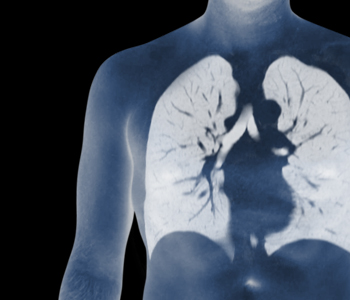
Lung
Inflammation, chronic diseases due to pollutants, tumours, pul-monary embolism
Abdominal organs
Tumours, inflammation, kidney stones, gallstones, intestinal ob-struction
Pelvis
Tumours, free fluid
Skeleton
Fractures, abrasive joint and spine diseases
Whole-body examination
Especially in the context of a “multiple myeloma” for measuring the tumour burden across the skeletal system
In most cases, a computed tomography will be performed as the result of unclear previous ex-ams, e.g. sonography or X-ray imaging, because the CT allows for a much better visualisation of anatomical structures and changes. In addition, post-calculations in all three dimensions can be carried out by taking a slice of the images, which leads to a considerable improvement in the diagnosis. In some cases, an MRI may be more useful than a CT, which might not be feasible due to a pacemaker, claustrophobia, or severe pain, however.
How is a computed tomography performed?
Equipment: Siemens Emotion 16, multi-line computer tomograph with progressive reduction of the radiation dose
A CT is an X-ray exam in which an X-ray tube revolves around the body while the table moves through the device. At the same time, images are taken in a spiral motion. This allows the recal-culation of slice images in 3 spatial directions.
You do not have to show up on an empty stomach. We require the functional values of your thyroid and kidney before the examination (these should be less than 2 months old), since in certain cases a contrast agent should not be administered in case of limited kidney function and thyroid hyperfunction.
For examinations of the abdominal area, you should take in 1/2 litre of contrast agent within 30 minutes before the start of the imaging procedure of the upper abdominal area or 1 litre within 60 minutes before the start of the imaging procedure of the entire abdominal area, which serves to delimit the gastrointestinal tract. During the examination, in many cases it becomes necessary to inject a contrast agent via a brachial vein. Thus, the blood flow of the organs and pathological processes that involved increased metabolism are captured. In turn, this allows for more sound diagnosis, since some of the changes can only be made visible by way of the contrast agent.
The contrast agent is usually very well tolerated, while the injection is associated with a feeling of warmth and a metallic taste in the mouth. Allergic reactions are possible in very rare cases. However, we can immediately treat a reaction with medication. If you have previously had an allergic reaction to the contrast agent, please let us know before the start of the examination.
The examination lasts about 5 to 10 minutes. The evaluation of the images takes between 10 and 20 min.
How To Get Here
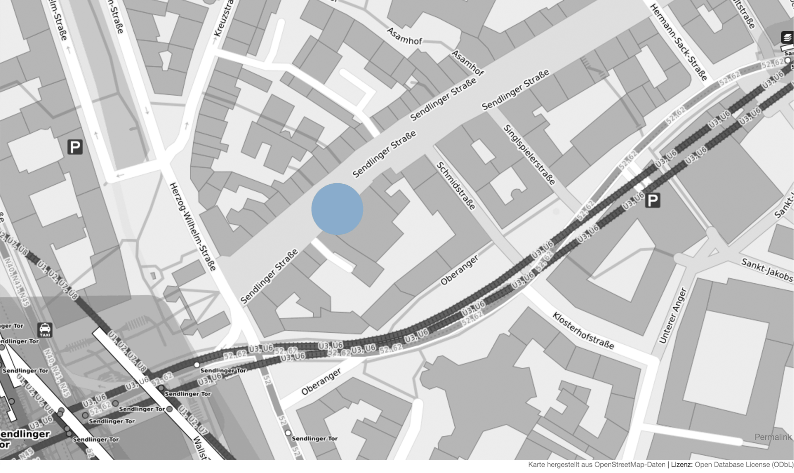
80331 Munich
Kontakt
Dr. med. Matthias BöheimDr. med. Andreas Vaitl
Dr. med. Johannes Vaitl
Phone 089 / 26 82 95
Fax 089 / 23 70 76 59
anmeldung@radiologie-sendlingerstrasse.de
Office Hours
Monday-Thursday8:00 - 18:00
Friday
8:00 - 14:00 Uhr